
The Dark Yet Fascinating History of Art, Science, & Medicine
- R.D. Ordovich-Clarkson

- Apr 21, 2025
- 5 min read
By Dr. Randall D. Ordovich-Clarkson, MD
The intersection of art, science, and medicine has a rich yet dark history that continues to shape both the way we study the human body and how we communicate our findings. I recently had the opportunity to share this fascinating journey with students and colleagues with the Henry Vandyke Carter Club’s first keynote speaker event, highlighting the role art has played in the advancement of medical knowledge (Ordovich-Clarkson, 2025). From the ancient anatomists of Hellenistic Alexandria to the groundbreaking work of Leonardo da Vinci, the fusion of artistic expression and scientific exploration has long been an integral part of our understanding of anatomy.
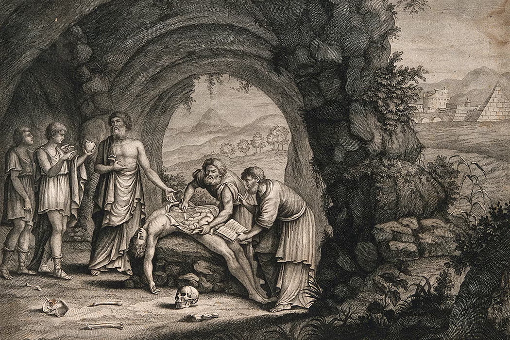
The Roots of Anatomical Art in Ancient Alexandria
Our journey begins in ancient Alexandria, where some of the earliest anatomical illustrations were created. Figures like Herophilus (335–280 BCE) and Erasistratus (c. 304–c. 250 BCE) made groundbreaking discoveries that formed the foundation of modern anatomy (Robb, 2024). Herophilus, often called the “Father of Anatomy,” was the first to perform dissections of human bodies, challenging the prevailing belief that the heart controlled the body, instead discovering that the brain was the true control center (Robb, 2024). His work, though lost in the infamous burning of the Library of Alexandria, set the stage for centuries of anatomical art that would follow.


Renaissance Masters: Da Vinci, Michelangelo, and the Revival of Anatomical Art
Fast forward to the Renaissance, a time when the study of anatomy was revived, largely due to the efforts of Leonardo da Vinci (1452–1519) and Michelangelo (1475–1564). These masters not only revolutionized art but also deepened our understanding of human anatomy. Leonardo, known for his meticulous anatomical drawings based on dissections, created some of the most accurate representations of the human body ever made. His famous Vitruvian Man remains a symbol of the connection between art and science, demonstrating the ideal human proportions based on anatomy (Lesso, 2024).
Similarly, Michelangelo’s sculptures, especially his depiction of the human form in the Sistine Chapel, show his deep understanding of anatomy. His detailed and accurate representations of the human body in art were informed by his study of cadavers, setting a new standard for anatomical accuracy in art (Lesso, 2024).
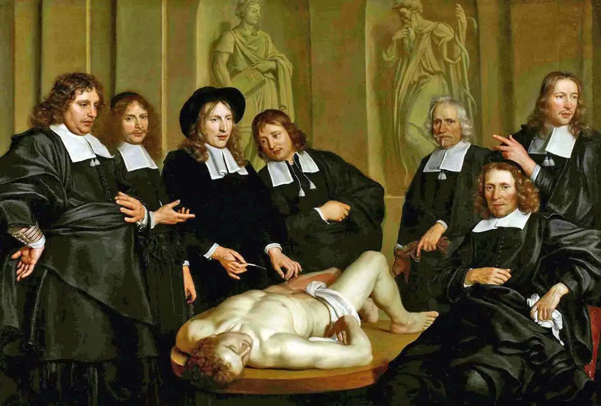
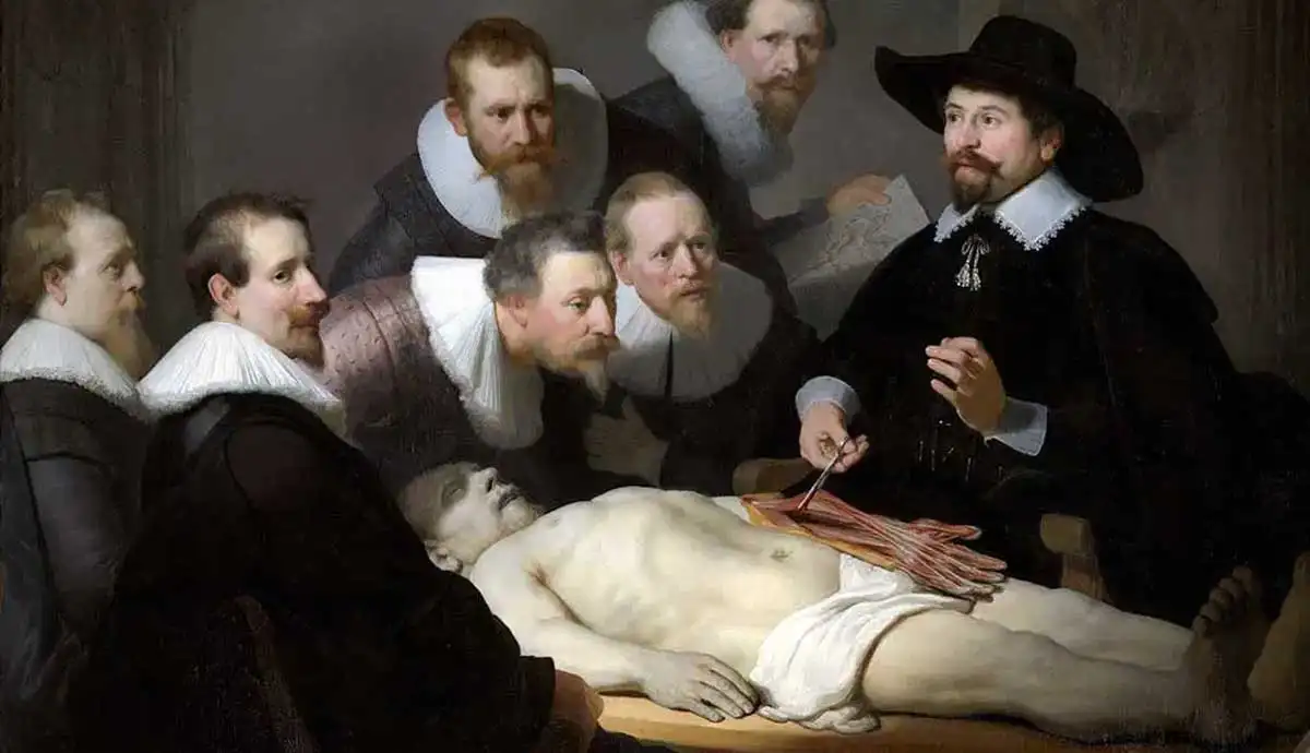
Andreas Vesalius: The Father of Modern Anatomy
One of the most significant figures in the history of anatomy is Andreas Vesalius (1514–1564), whose work De Humani Corporis Fabrica (On the Fabric of the Human Body) forever changed the field. Published in 1543, Vesalius’ text provided detailed illustrations of the human body based on direct dissections, a major shift from the reliance on ancient texts. His collaboration with the artist Jan Steven van Calcar produced some of the most famous anatomical illustrations of the time (Shelbourn, 2006). Vesalius’ work not only advanced medical knowledge but also established anatomy as a scientific discipline rooted in empirical observation.


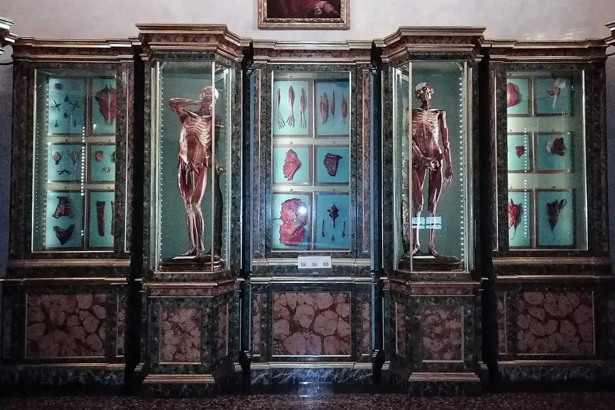
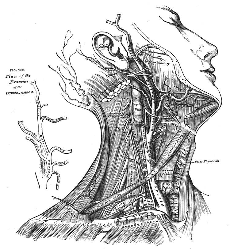
Wax Anatomical Models: Bridging Art and Medicine
During the 17th to 19th centuries, as dissection became a more formal part of medical education, anatomical art took on new forms with the creation of wax anatomical models. These lifelike representations were used to teach students about the intricacies of human anatomy without the need for preserving actual cadavers. Museums like La Specola in Florence and The Mütter Museum in Philadelphia house collections of these stunningly detailed models, many of which were painstakingly crafted by skilled artists to depict muscles, veins, and organs in realistic detail (Jacobson, 2016). These models were not only educational tools but also beautiful works of art, bridging the gap between scientific learning and artistic expression.


Modern Anatomical Art: From Frank Netter to Gunther von Hagens
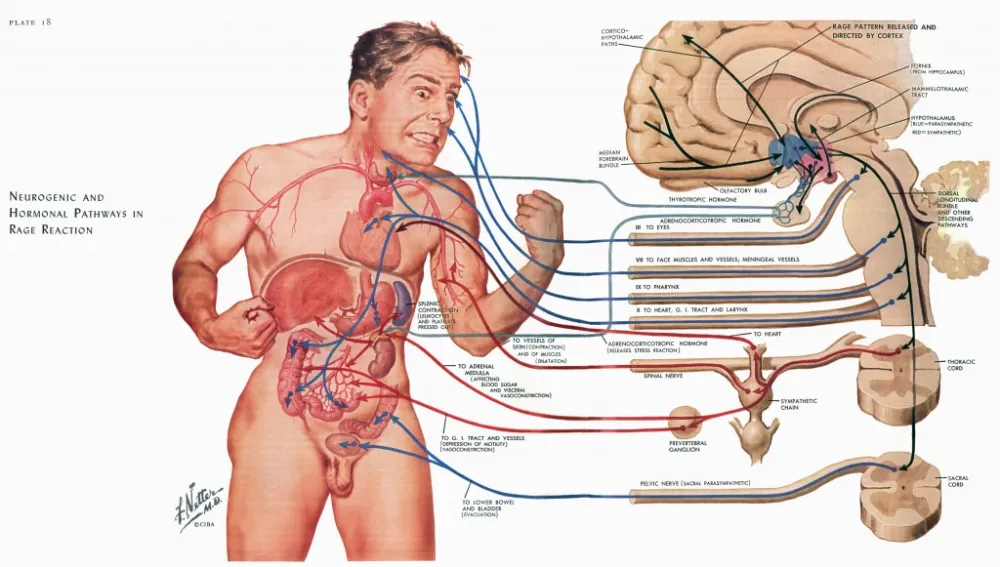
In the 20th century, the legacy of anatomical art continued with the work of Dr. Frank Netter (1906–1991), whose medical illustrations became standard references in textbooks and clinical settings. Netter’s work is often compared to that of Norman Rockwell, as his ability to convey the human body with both scientific precision and artistic flair elevated his illustrations to iconic status. His drawings continue to be used in modern medical education and patient education (Netter, 1991).
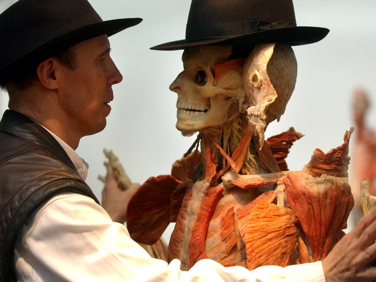
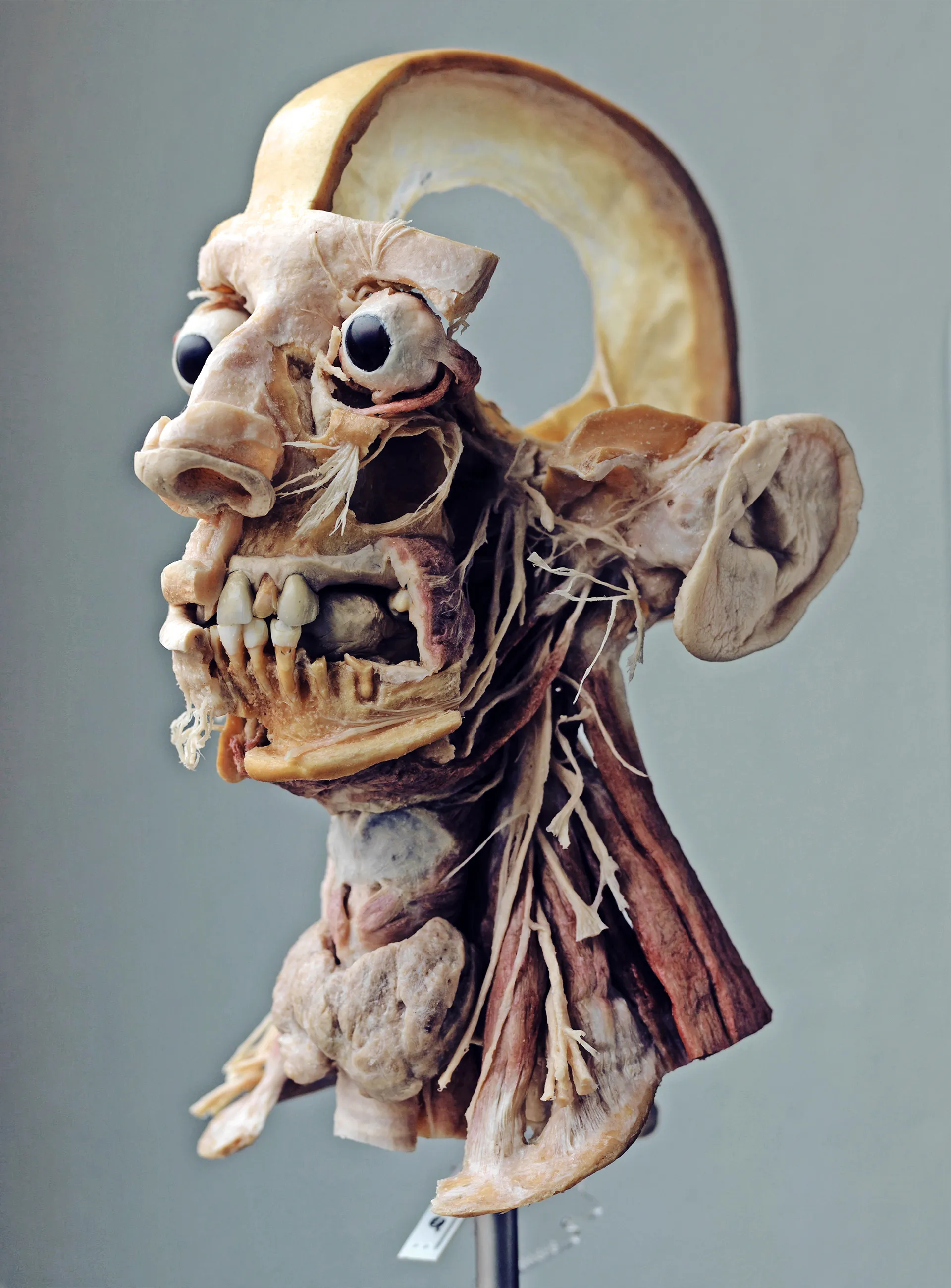
However, as we entered the modern era, the development of new technologies introduced new ways to study and display human anatomy. Gunther von Hagens, a German anatomist, pushed the boundaries of anatomical art with his creation of plastination, a method of preserving human bodies by replacing water and fat with plastics. His Body Worlds exhibition, which showcases plastinated bodies, has traveled the world, offering audiences a tangible, 3D view of human anatomy. Although his work has sparked ethical debates, particularly regarding the use of “unclaimed” human bodies, it has undeniably advanced public understanding of human anatomy in ways that were previously unimaginable (von Hagens, 1995).


The Continuing Role of Art in Science and Medicine
Today, the intersection of art, science, and medicine continues to thrive. While digital imaging and 3D modeling have revolutionized the way we study anatomy, the value of artistic representation in medical education remains profound. Artistic works—whether traditional illustrations, wax models, or digital images—continue to help students and professionals visualize complex anatomical structures and understand the human body in ways that cannot be achieved through text alone. As I discussed in my talk, the integration of art in medical education enriches our learning experiences, fosters creativity, and enhances our ability to communicate complex concepts.
Conclusion: A Timeless Fusion
Art has been integral to the study and understanding of the human body for centuries, and it will undoubtedly continue to play a vital role in both medical education and healthcare. From the early anatomical illustrations of ancient Alexandria to the groundbreaking work of Leonardo da Vinci, Vesalius, and Netter, to the modern techniques of plastination and digital imaging, the fusion of art and science has been a constant force in the advancement of medical knowledge. As we move forward, it is crucial to recognize the importance of this connection and continue to explore the ways in which art can inform and enhance our understanding of the human form.
References:
Case Western Reserve University. (2014, May 22). Under Your Skin: the Anatomy Artwork of H.V. Carter. Dittrick Medical History Center. https://artsci.case.edu/dittrick/2014/05/22/under-your-skin-the-anatomy-artwork-of-h-v-carter/
Bansal, D. (2017). Digital archive of antique wax figures becomes a teaching tool. News Center. https://med.stanford.edu/news/all-news/2017/04/digital-archive-of-antique-wax-figures-becomes-teaching-tool.html
Hussey, K. D. (2017) “Seen and Unseen: The Representation of Visible and Hidden Disease in the Waxworks of Joseph Towne at the Gordon Museum”, 19: Interdisciplinary Studies in the Long Nineteenth Century.(24). doi: https://doi.org/10.16995/ntn.787
Jacobson, M. M. (2016). 18 Places To See Uncanny Specimens of Wax Anatomy. Atlas Obscura. https://www.atlasobscura.com/lists/18-places-to-see-uncanny-specimens-of-wax-anatomy
Lesso, R. (2024, September 21). How Did Artists Learn About Anatomy During the Renaissance? TheCollector. https://www.thecollector.com/artists-learn-renaissance-anatomy/
NYU. (n.d.). Armamentarium Chirurgicum. NYU Dentistry. https://dental.nyu.edu/aboutus/rare-book-collection/17-c/johanni-scultetus.html
Ordovich-Clarkson, R. D. (2025, April 3). A Brief History of Art, Science, & Medicine [Conference presentation]. Exploring the Intersection of Science and Creativity, Grand Canyon University, Phoenix, AZ. http://dx.doi.org/10.13140/RG.2.2.11631.55202
Robb, D. (2024, November 23). The Anatomists of Ancient Alexandria - JSTOR Daily. JSTOR Daily. https://daily.jstor.org/the-anatomists-of-ancient-alexandria/
Netter, F. (1991). Netter’s Atlas of Human Anatomy. Elsevier.
Shelbourn, C. (2006). Bringing the Skeletons out of the Closet: The Law and Human Remains in Art, Archaeology and Museum Collections. Art Antiquity & Law, 11, 179–198.
von Hagens, G. (1995). Plastination: The New Anatomy Art. Body Worlds Exhibition.


Comments